
Restriction Enzyme Digestion
Preparation of DNA for traditional cloning methods is dependent upon restriction enzyme digestion to generate compatible ends capable of being ligated together. The DNA to be cloned can vary widely, from genomic DNA extracted from a pure bacterial culture or a mixed population, to a previously cloned gene that needs to be moved from one vector to another (subcloning). Restriction enzymes can also be used to generate compatible ends on PCR products. In all cases, one or more restriction enzymes are used to digest the DNA resulting in either non-directional or directional insertion into the compatible plasmid.
Genomic DNA, regardless of the source, is typically digested with restriction enzymes that recognize 6-8 consecutive bases, as these recognition sites occur less frequently in the genome than 4-base sites, and result in larger DNA fragments. The desired insert size for the clone library determines which enzymes are selected, as well as the digestion conditions. Most often, a serial dilution of the selected restriction enzyme(s) is used to digest the starting material and the desired insert size range is isolated by electrophoresis followed by gel extraction of the DNA. This method of preparation provides DNA fragments of the desired size with ends compatible to the selected vector DNA.
Restriction enzymes, first described in 1971, are bacterially derived enzymes that cleave DNA. Evolutionarily, restriction enzymes arose as a bacterial self-defense mechanism; the genomes of invading organisms would be degraded, leading to an inability to replicate. Type II restriction enzymes generally recognize an inverted repeat palindrome. This recognition site structure leads to a symmetrical cleavage of both DNA strands and results in either blunt- or sticky-ends of the digested DNA. Blunt ends are universally compatible with other blunt-ended DNA and possess a 5’ phosphate group to promote ligation. Sticky ends, on the other hand, are stretches of single-stranded DNA that is capable of self-ligation or ligation with a complementary region of DNA from another molecule or organism.
Use Enzyme Finder to select restriction enzymes by name, sequence, overhang or type.
Choose Type:
- Removal of Single-Stranded Extension Protocol using Mung Bean Nuclease (M0250)
- Standard Digest Using RE-Mix®
- Double Digest Protocol using Two RE-Mix® Enzymes
- Optimizing Restriction Endonuclease Reactions
- Protocol for Cre Recombinase (M0298)
- Double Digest Protocol using One RE-Mix and One Standard Restriction Enzyme
- Protocol for Glucosylation and digestion of Genomic DNA using AbaSI (#R0665)
- Protocol for Direct Digestion of gDNA during droplet digital PCR (ddPCR)
- Double Digest Protocol with Standard Restriction Enzymes
-
Restriction Enzymes at NEB: Over 30 years of Innovation
-
Restriction Endonucleases: Molecular Cloning and Beyond
-
Type II Restriction Enzymes: What You Need to Know | NEB
Read about Type II restriction enzymes and the distinguishing properties of the four principle subtypes.
-
A Modern Day Gene Genie Sir Richard Roberts on Rebase
-
Whole genome assembly from next generation sequencing data using restriction and nicking enzymes in optical mapping and proximity-based ligation strategies
High throughput sequencing methods have revolutionized genomic analysis by producing millions of sequence reads from an organism’s DNA at an ever decreasing cost.
- Molecular Cloning Technical Guide
- Recleavable Filled-in 5' Overhangs
- Alphabetized List of Recognition Sequences
- Compatible Cohesive Ends and Generation of New Restriction Sites
- Dam-Dcm and CpG Methylation
- Recleavable Blunt Ends
- Why Choose Recombinant Enzymes?
- Cleavage Of Supercoiled DNA
- Isoelectric Points (pI) for Restriction Enzymes
- Enzymes with Nonpalindromic Sequences
- Enzymes with Multiple Recognition Sequences
- Frequencies of Restriction Sites
- Interrupted Palindromes
- Isoschizomers
- Troubleshooting Guide for Cloning
- Restriction Enzyme Troubleshooting Guide
- Activity at 37°C for Restriction Enzymes with Alternate Incubation Temperatures
- Restriction Endonucleases - Survival in a Reaction
- NEBuffer Activity/Performance Chart with Restriction Enzymes
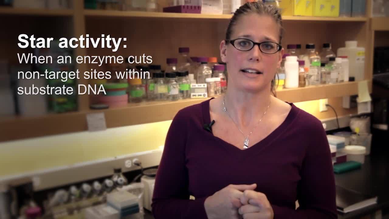
- Star Activity
- Digestion of Agarose-Embedded DNA: Info for Specific Enzymes
- Alteration of Apparent Recognition Specificities Using Methylases
- Cleavage Close to the End of DNA Fragments
- Traditional Cloning Quick Guide
- Restriction Enzymes for Droplet Digital PCR (ddPCR)
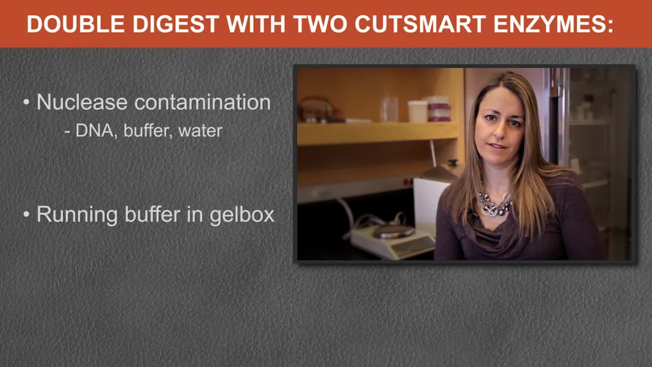
- Double Digests
- Optimizing Restriction Endonuclease Reactions
- Restriction Enzyme Tips
- Heat Inactivation
- Site Preferences
Feature Articles
Brochures
Selection Tools
Troubleshooting Guides
Usage Guidelines
- Fu YB, Peterson G. W., Dong Y (2016) Increasing Genome Sampling and Improving SNP Genotyping for Genotyping-by-Sequencing with New Combinations of Restriction Enzymes G3 (Bethesda); 6:4, 845-846. PubMedID: 26818077
- Shah, S., Sanchez, J., Stewart, A., et al. (2015) Probing the Run-On Oligomer of Activated SgrAI Bound to DNA PLoS One; 10(4), PubMedID: 25880668, DOI: 10.1371/journal.pone.0124783.
- Loenen, W.A., Raleigh, E.A. (2014) The other face of restriction: modification-dependent enzymes. Nucleic Acids Res; 42, 56-69. PubMedID: 23990325, DOI: doi: 10.1093/nar/gkt847
Products and content are covered by one or more patents, trademarks and/or copyrights owned or controlled by New England Biolabs, Inc (NEB). The use of trademark symbols does not necessarily indicate that the name is trademarked in the country where it is being read; it indicates where the content was originally developed. All other trademarks are the property of their respective owners. The use of this product may require the buyer to obtain additional third-party intellectual property rights for certain applications. For more information, please email busdev@neb.com.
This product is intended for research purposes only. This product is not intended to be used for therapeutic or diagnostic purposes in humans or animals.
Choose Product:
Subcloning requires the use of 1-2 restriction enzymes that cut immediately outside the insert fragment without cutting within the insert itself. Restriction enzymes that have a recognition site within the multiple cloning site (MCS) are commonly used since they do not cut elsewhere in the vector DNA and typically produce two easily resolved DNA fragments. The gene of interest is most commonly subcloned into an expression vector for improved protein expression and/or addition of a purification tag. In this case, it is essential that the gene be inserted in the correct orientation and in frame with the transcription promoter.
The Polymerase Chain Reaction (PCR) is commonly used to amplify a gene or DNA fragment of interest, from any source of DNA, to be cloned. In order to generate compatible ends, it is common to add restriction sites to the 5’ end of both PCR primers. When adding restriction sites to a PCR primer, it is recommended to include 6 bases between the recognition site and the 5’ end of the primer. These additional bases provide sufficient DNA for the restriction enzyme to bind the recognition site and cut efficiently. When selecting a restriction site(s) to add to the primers, it is important to determine which site(s) will be compatible with your selected vector, whether directional cloning is desired and, most importantly, confirm that the recognition site(s) does not occur within the gene or DNA fragment.
-
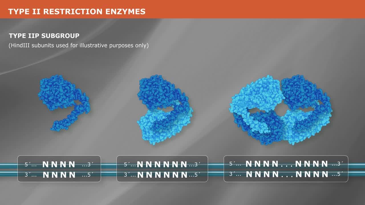
What is a Type II Restriction Enzyme?
-
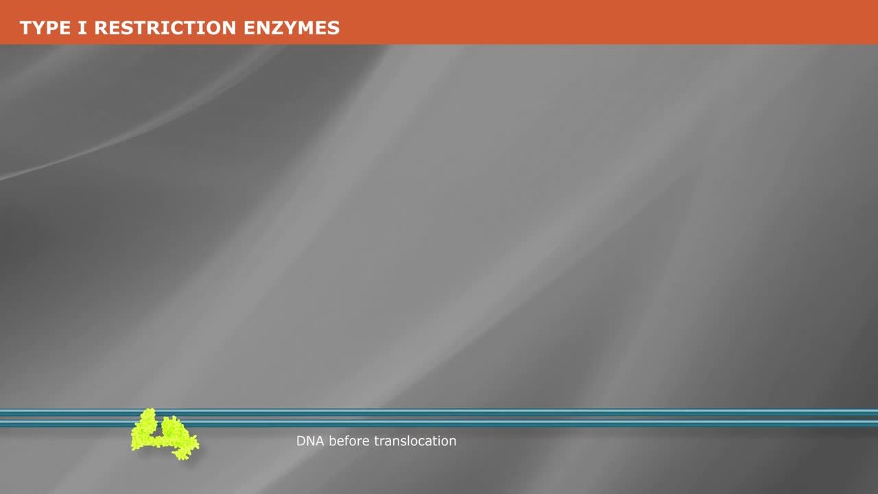
What is a Type I Restriction Enzyme?
-
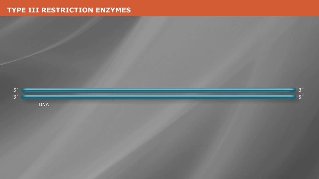
What is a Type III Restriction Enzyme?
-

Cloning With Restriction Enzymes
-
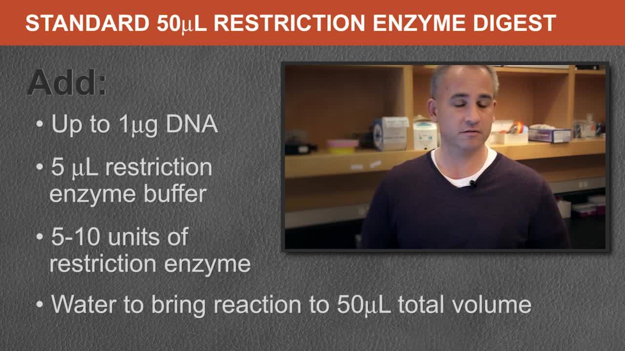
Standard Protocol for Restriction Enzyme Digests
-

Why is My Restriction Enzyme Not Cutting DNA?
-
Restriction Enzyme Digest Problem: Too Many DNA Bands
-
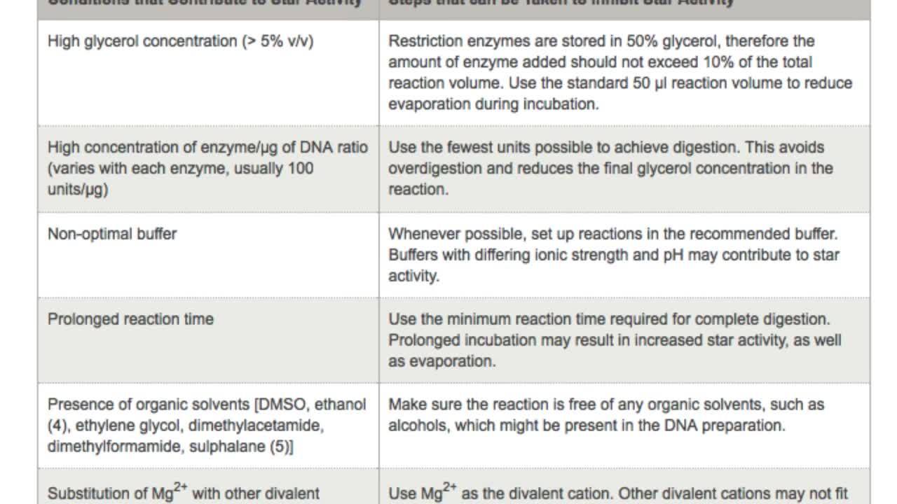
What is Restriction Enzyme Star Activity?
-

Reduce Star Activity with High-Fidelity Restriction Enzymes
-
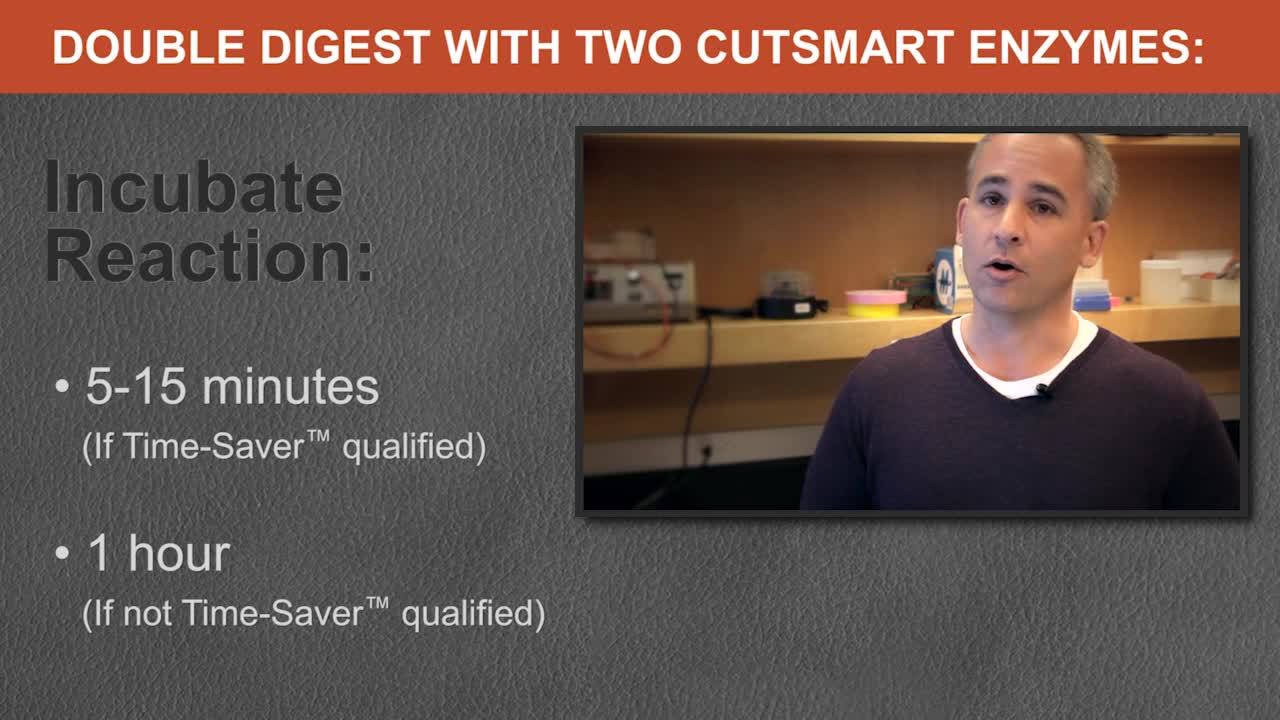
NEB® Restriction Enzyme Double Digest Protocol
-

Restriction Enzyme Digest Protocol: Cutting Close to DNA End
-
Restriction Enzyme Digestion Problem: DNA Smear on Agarose Gel
-

Restriction Enzymes in Golden Gate Assembly
-

Restriction Enzymes in Optical Mapping
