
RNase 4 Supports Precise Characterization of mRNA 5´ Cap Structures
Posted on Monday, September 14, 2026
By
Topic: Tips for the lab
Analytic tools to characterize complex messenger RNA (mRNA) sequences are crucial for mRNA-based therapeutics and vaccines. In a July 2026 application note, New England Biolabs researchers highlight an mRNA characterization protocol using RNase 4 to generate mRNA 5´ cap oligonucleotide sequences for analysis via liquid chromatography tandem mass spectrometry (LC-MS). This enzymatic digestion is required when using LC-MS for lengthy mRNAs.
For this protocol, researchers obtained National Institute of Standards and Technology Research-Grade Test Material 10202 (NIST RGTM 10202) to support the assessment of mRNA Critical Quality Attributes (CQAs). As a result, the specific protocol amounts referenced in the application note have been optimized for NIST RGTM 10202 mRNA.
In addition to an mRNA characterization protocol, the application note provides recommendations for streptavidin magnetic bead enrichment.
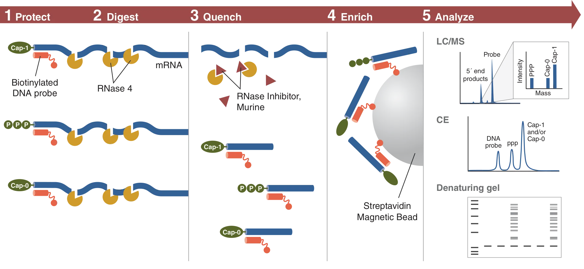
Overview of an mRNA characterization protocol using RNase 4 to generate mRNA 5´ cap oligonucleotide sequences, which can be analyzed using LC-MS, capillary electrophoresis (CE), or gel-based separation under denaturing conditions.
A strategic protocol for the site-directed RNase 4 cleavage of mRNA 5´ ends & enrichment with protective biotin-DNA probe
RNase 4 is an RNA endonuclease that cleaves 3´ of uridine in uridine-purine sequences. It allows for more targeted RNA digestion compared to single-nucleotide specific RNases and tolerates uridine base modifications. However, RNase 4 is specific for single-stranded RNA (ssRNA), so a protective biotin-DNA probe is required to protect an RNA region from digestion.

RNase 4 cuts RNA at U/A and U/G.
After hybridization with the biotin-DNA probe, the mRNA 5' end is protected from RNase 4 digestion, and cleavage occurs at the next available U/A or U/G site. The reaction is then quenched with an RNase Inhibitor. The probe-hybridized mRNA 5´ product can be captured using streptavidin magnetic beads and analyzed using LC-MS, capillary electrophoresis (CE), or gel-based separation under denaturing conditions.
Recommendations for magnetic steptavidin beads
Our scientists found that using a higher volume of beads for RNA:DNA duplex capture increases the relative proportion of RNA-specific signal by LC-MS, independent of whether biotin or desthiobiotin was used and of probe conjugation. They also noted better performance when hydrophobic streptavidin beads were used compared to hydrophilic beads.
Effectively quantify mixtures of 5´ cap mRNA material and assess mRNA CQAs
To learn more about how NEB researchers conducted their RNase 4-based workflow for mRNA 5´ cap characterization, including RNase 4 unit optimization, supporting figures, and more, refer to our application note, “RNase 4 Enables Targeted Isolation and Quantitative Analysis of mRNA 5´ Cap Structures.”
Don’t miss out on our latest NEBinspired blog releases!
- Sign up to receive our e-newsletter
- Download your favorite feed reader and subscribe to our RSS feed
Be a part of NEBinspired! Submit your idea to have it featured in our blog.