
In vivo Imaging
The dynamic real-time localization and translocation of molecules in living cells is an integral aspect of cellular function. Scientists are pioneering new applications for in vivo cellular and molecular imaging. These applications are based on genetic engineering advances and improved imaging technologies. In vivo imaging has become quantifiable, highly sensitive and amenable to high-throughput study design. The high sensitivity and utility of in vivo imaging, is exemplified by use of the Fluorescence Resonance Energy Transfer (FRET) method, which quantitates enzyme activity, protein-protein interactions and second messenger dynamics. Optical in vivo imaging methods are being applied in pre-clinical research studies. Real time in vivo imaging studies in whole animals have been used to track cell-based therapies, and monitor response to chemotherapies (1).
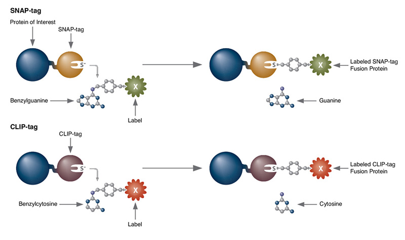
Fluorescence based recombinant systems are attractive molecular imaging tools because of their advantages for in vivo studies. Both bioluminescence reporters and fluorescence labeling systems offer relatively easy transition from in vitro, to living cells, tissues or small whole animal analysis (2). These protein labeling and reporter systems are non-invasive and allow direct analysis of cellular and molecular dynamics in real time. Some examples of bioluminescent reporters useful for in vivo imaging are enhanced GFP, FAP, Gaussia Luciferase (GLuc) and Cypridina Luciferase (Cluc). Protein labeling systems such as Tetra-Cys, SNAP-tag® and CLIP-tag™ can also be used for in vivo techniques. Imaging in living animals is currently best accomplished by PET reporters for scanning with short-lived radioactive tracers. Multimodality reporter gene systems have also been designed to permit in vivo imaging by multiple detection methods (3). For any fluorescence imaging application, fluorophore selection for native target activities and photostability should be confirmed in heterogeneous cellular environments.
References
- Kircher, M.F.,Gambhir, S.S., Grimm, J. (2011) Nature Rev. Clinical Oncology, 8, 677-88. PMID: 21946842
- Keyaerts, M., Caveliers, V., and Lahoutte, T. (2012) Trends in Molecular Medicine. Epub 1-9. PMID: 22321645
- Ray, P. (2011) J. Biosci. 36: 499–504. PMID: 21799261
SNAP-tag® is a registered trademark of New England Biolabs, Inc.
Choose Type:
- Cellular Labeling (E9100)
- Cellular Labeling (E9230)
- Cellular Labeling (S9103)
- Protocol for Cellular Labeling with SNAP-Cell® Oregon Green® (NEB #S9104)
- SNAP-Cell® TMR-Star Cellular Labeling (NEB #S9105)
- Cellular Labeling (S9107)
- Cellular Labeling (S9109)
- Cellular Labeling (S9110)
- Cellular Labeling (S9112)
- Cellular Labeling (S9124)
- Cellular Labeling (S9129)
- Cellular Labeling (S9132)
- Cellular Labeling (S9134)
- Cellular Labeling with SNAP-Surface® Alexa Fluor® 647 (NEB #S9136)
- Cellular Labeling (S9217)
- Cellular Labeling (S9219)
- Cellular Labeling (S9232)
- Cellular Labeling (S9233)
- Cellular Labeling (S9234)
- Cloning of CLIP-tag Fusions in pCLIPf (N9215)
- CoA 488 (S9348)
- Expression of CLIP-tag Fusions (N9215)
- Labeling of Proteins in vitro (S9110)
- Labeling of Proteins in vitro (S9103)
- Labeling of Proteins in vitro (S9104)
- Labeling of Proteins in vitro (S9105)
- Labeling of Proteins in vitro (S9106)
- Labeling of Proteins in vitro (S9107)
- Labeling of Proteins in vitro (S9109)
- Labeling of Proteins in vitro (S9143)
- Labeling of Proteins in vitro (S9217)
- Labeling of Proteins in vitro (S9219)
- Labeling of Proteins in vitro (S9220)
- Labeling of Proteins in vitro (S9221)
- Labeling of Proteins in vitro (S9232)
- Labeling of Proteins in vitro (S9233)
- Labeling of Proteins in vitro (S9234)
- Instructions for Labeling of Proteins in vitro (S9348)
- Labeling of Proteins in Solution (E9230)
- Labeling on the Surface of Cells (S9349)
- Labeling on the Surface of Cells (S9350)
- Labeling Proteins in vitro (S9112)
- Labeling Proteins in vitro (S9124)
- Labeling Proteins in vitro (S9129)
- Labeling Proteins in vitro (S9132)
- Labeling Proteins in vitro (S9134)
- Labeling Proteins in vitro (S9136)
- Use of SNAP-Cell Block with SNAP-Cell Substrates (E9100)
- Use of CLIP-Cell Block with CLIP-Cell Substrates (E9230)
- Use with CLIP-tag substrates (S9220)
- View the video "Fluorescent Labeling of COS-7 Expressing SNAP-tag Fusion Proteins for Live Cell Imaging" in the Journal of Visualized Experiments (JoVE)
- Cellular Labeling (E9120)
- Instructions for Cellular Labeling (E9200)
- Labeling Proteins in vitro (E9120)
- Labeling Proteins in vitro (E9200)
- Cloning of SNAP-tag Fusions in pSNAPf (N9183)
- Cloning of SNAP-tag Fusions in pSNAP-tag(T7)-2 (N9181)
- Expression of SNAP-tag Fusions (N9181)
- Expression of SNAPf Fusions (N9183)
- Labeling of Proteins in vitro (S9349)
- Labeling of Proteins in vitro (S9350)
- Cellular Labeling (S9221)
- Labeling of Proteins in vitro (E9100)
- Use with SNAP-Surface substrates (S9143)
- Use with SNAP-Cell Substrates (S9106)
- Cellular Labeling (S9159)
- Labeling of Proteins in vitro (S9159)
- SNAP-Cell® 647-SiR Cellular Labeling (NEB #S9102)
- Labeling of Proteins in vitro (S9102)
- Reuter, W.H., Masuch, T., Ke, N., Lenon, M., Radzinski, M., Van Loi, V., Ren, G., Riggs, P., Antelmann, H., Reichmann, D., Leichert, L.I., Berkmen, M (2019) Utilizing redox-sensitive GFP fusions to detect in vivo redox changes in a genetically engineered prokaryote Redox Biol; 26, 101280. PubMedID: 31450103, DOI: 10.1016/j.redox.2019.101280
- Maffei, M., Morelli, C., Graham, E., Patriarca, S., Donzelli, L., Doleschall, B., de Castro, Reis, F., Nocchi, L., Chadick, C.H., Reymond, L., Correa, I.R., Jr., Johnsson, K., Hackett, J.A., Heppenstall, P.A (2019) A ligand based system for receptor specific delivery of proteins Sci Rep; 9(1), 19214.. PubMedID: 31844114, DOI: 10.1038/s41598-019-55797-1
- Ke, N., Landgraf, D., Paulsson, J. and Berkmen, M. (2016) Visualization of Periplasmic and Cytoplasmic Proteins with a Self-Labeling Protein Tag. J Bacteriol; Jan 19;198(7), 1035-43. PubMedID: 26787765
- Clone and express once, then use with a variety of substrates
- Non-toxic to living cells
- Wide selection of fluorescent substrates
- Highly specific covalent labeling
- Simultaneous dual labeling
- Simultaneous dual protein labeling inside live cells
- Protein localization and translocation
- Pulse-chase experiments
- Receptor internalization studies
- Selective cell surface labeling
- Protein pull-down assays
- Protein detection in SDS-PAGE
- Flow cytometry
- High throughput binding assays in microtiter plates
- Biosensor interaction experiments
- FRET-based binding assays
- Single molecule labeling
- Super-resolution microscopy

Products and content are covered by one or more patents, trademarks and/or copyrights owned or controlled by New England Biolabs, Inc (NEB). The use of trademark symbols does not necessarily indicate that the name is trademarked in the country where it is being read; it indicates where the content was originally developed. All other trademarks are the property of their respective owners. The use of this product may require the buyer to obtain additional third-party intellectual property rights for certain applications. For more information, please email busdev@neb.com.
This product is intended for research purposes only. This product is not intended to be used for therapeutic or diagnostic purposes in humans or animals.
