How do restriction enzymes interact with DNA?

Script
Geoffrey Wilson:
The restriction enzyme free in the cell is a fairly sort of open structure. A lot of these restriction enzymes act as dimers. That is to say, there are two identical sub-units that bind to each other and they have an open structure that'll encounter a DNA molecule and close around it. And, that creates either a sort of screw twist or a cylinder that'll then slide along DNA. It'll spin down the DNA. As it's spinning down the DNA, it's encountering DNA sequences the whole time and, for the most part, those sequences will be the wrong sequence and they won't attach to them.
But, just now and again, when they encounter the right sequence, the shape will be right, the electrostatics will be right, and the enzyme will bind to that sequence. It stops. It remains bound and they can remain bound for very long periods of time. But normally, what would happen would be they would bind and then immediately catalyze reaction, within a second or two. They would cleave both strands of the DNA, and having done that, then their substrate is destroyed. Then they would fall off and they would go into the open confirmation again, reattach, and then continue sliding.
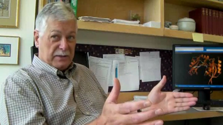
This is the structure of a restriction enzyme called Mva I. What you'll notice is that this has a shape somewhat like a hand. You could imagine this to be a thumb and these could be fingers. And, it has this shape because this is going to grasp the DNA molecule. It's gonna grasp the molecule. Once it's grasped it, it's gonna slide along it until it encounters its recognition sequence. Pink is without the DNA so this is the free enzyme on its own, as it would exist in the cell, when it was about to encounter the DNA molecule but hadn't.
Now, in orange we're looking at the structure of precisely the same protein when it has encountered DNA. The structure changes when it attaches to DNA.
Notice this loop here and this loop here. Both of them closed. So, in orange now, we have the structure of exactly the same protein, but now this is bound to DNA. You notice tremendous movements of regard. This enzyme has wrapped itself almost completely around the DNA.
This is the DNA molecule that the enzyme has crystallized with. This has the sequence CC(A or T)GG which is the recognition sequence of this enzyme. Now, I put in the enzyme and you'll notice that the enzyme is intimately associated with the majority of the DNA.
This is the surface structure now of the enzyme when it's bound to DNA. The protein has almost completely encircled the DNA. There's just a small groove in here which is following the contour of the double helix and the protein follows the helical turns, and almost completely encloses the DNA.
Related Videos
-

What are Restriction Enzymes? -

Standard Protocol for Restriction Enzyme Digests -

Discovering New Restriction Enzymes

