Fluorescent Labeling of COS-7 Expressing SNAP-TAG® Fusion Proteins for Live Cell Imaging
Script
Narrator:
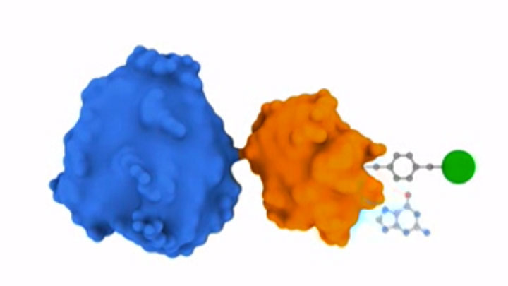
SNAP- and CLIP-tag protein labeling systems enable the specific covalent attachment of virtually any molecule to a protein of interest. The gene of interest can be cloned into the vector on either side of the tag. Expression of the cloned gene results in a SNAP-tag fusion protein.
The SNAP-tag substrate consists of two parts: the benzylguanine, which is recognized by the SNAP-tag, and the functional group. The functional group may be biotin, a bead, or a fluorescent group, which are available in a variety of colors.
During the labeling reaction, the substituted benzyl group covalently attaches to the SNAP-tag, releasing guanine. If a fluorophore is coupled to the desired protein, the label fluoresces, permitting visualization of the protein in living or fixed cells.
Chris Provost:
Hi, I'm Chris Provost from New England Biolabs. Today we will show you a procedure for the fluorescent imaging of live COS-7 cells expressing SNAP-tag fusion proteins. We use this procedure in our laboratory to study where proteins are localized in cells. So let's get started.
Narrator:
To prepare COS-7 cells for transfection, trypsinize them according to standard protocols and add 50 to 100 microliters of cells to six milliliters of complete DMEM medium. Mix the cell suspension by pipetting up and down several times, then aliquot 300 microliters of trypsinized cell suspension to each well of a sterile eight-chamber Lab-Tek ii Chambered Coverglass. Incubate samples at 37 degrees Celsius, 5% carbon dioxide overnight.
On the following day, check the cell culture chambers for cell density and health. Label the appropriate number of microfuge tubes for the transfection complex mixtures.
In a laminar flow hood, set up the transfection complex mixtures. For each reaction use 0.3 micrograms of SNAP-fusion plasmid DNA, diluted in serum-free medium, and one microliter each of the TransPass D2 and TransPass V transfection reagents.
Control plasmids are available to assay for the efficacy of the transfection reagents. We also recommend including a mock transfected sample and/or an untransfected sample as negative controls.
For each complex mixture, first mix the serum-free DMEM with the appropriate plasmid DNA, then add the TransPass D2 transfection reagent, followed by the TransPass V transfection reagent. Mix the components well by pipetting up and down gently several times after the addition of each reagent, and incubate the reactions at room temperature for 30 minutes.
During the incubation, wash cells once with 600 microliters of complete DMEM and add 450 microliters of complete DMEM to each sample. When the incubation is complete, add 50 microliters of the transfection complex mixture to each sample, slowly and in a dropwise fashion. Incubate the samples overnight at 37 degrees Celsius, 5% carbon dioxide.
The next day, the transfected COS-7 cells can be labeled with the SNAP-Cell TMR-Star substrate. To begin this procedure, add 50 microliters of DMSO to the SNAP-Cell TMR-Star substrate and pipette up and down. Mix by vortexing for one minute. Next, carefully remove the transfection complex media from each sample by vacuum suction. Wash cells twice with 600 microliters of complete DMEM, then replace the media in each well with 600 microliters of complete DMEM. Return the samples to the incubator.
Mix dye labeling media by adding 995 microliters of complete DMEM and five microliters of 0.6 millimolar SNAP-Cell TMR-Star substrate. Pipette up and down several times to mix after the addition of substrate. The final concentration of SNAP-Cell TMR-Star should be 3 micromolar. Carefully remove growth media from the samples and add 200 microliters of dye labeling media to each well. Incubate samples at 37 degrees Celsius, 5% carbon dioxide for 30 minutes.
It is at this step that the benzyl group on the TMR substrate will covalently link to the SNAP-tag and release guanine.
While the samples are incubating, prepare Hoechst solution for nuclear staining of samples. Mix one microliter of Hoechst 33342 trihydrochloride trihydrate and 10 milliliters of complete DMEM. When the incubation is complete, remove the dye labeling media from all samples and add 600 microliters of the dilute Hoechst solution to each sample. Incubate samples at 37 degrees Celsius, 5% carbon dioxide for five minutes.
After five minutes, remove the dilute Hoechst solution from each sample and wash the samples three times with 600 microliters of complete DMEM. After the final wash, add 600 microliters of complete DMEM to each sample. Incubate the samples at 37 degrees Celsius, 5% carbon dioxide for an additional 30 minutes to allow unincorporated fluorophor to diffuse out of the cells, which is required for intracellular labeling.
After 30 minutes, replace the media on the SNAP-Cell samples one last time with 600 microliters of complete DMEM. The cells are now ready for imaging, using a Zeiss ApoTome Axiovert 200M fluorescent microscope.
Here are some representative results of fluorescent imaging, using standard fluorescent microscopy of live COS-7 cells that are expressing SNAP-tag fusions.
This first image shows live COS-7 cells expressing pSNAP-H2B, labeled with SNAP-Cell TMR-Star. The pSNAP-H2B construct was generated using the pSNAP-tag (m) Vector. The cells were labeled with SNAP-Cell TMR-Star for 30 minutes and counterstained with Hoechst 33342 for the nuclei. The pink fluorescence demonstrates the overlay of red fluorescence where histone protein is present in addition to the blue fluorescence, indicating the nucleus. The expression of the H2B protein is clearly and easily identified.
These next two images show live COS-7 cells transiently transfected with pSNAP-ADR Beta2. Cells were labeled with SNAP-Cell TMR-Star and counterstained with Hoechst 33342. The pSNAP-ADR Beta2 construct was generated using the pSNAP-tag (m) Vector. The cells were labeled with SNAP-Cell TMR-Star for 30 minutes and counterstained with Hoechst 33342 for nuclei. The red fluorescence indicates the presence of cell-surface receptor protein ADR Beta2, while blue fluorescence identifies the nucleus.
Chris Provost:
We've just shown you how to perform fluorescent imaging of live COS-7 cells expressing SNAP-tag fusion proteins. When doing this procedure, it's important to remember to add the transfection complex slowly and in a dropwise fashion and to store the SNAP-tag substrate at minus 20 degrees Celsius in the dark when not in use.
So that's it. Thanks for watching, and good luck with your experiments.
Related Videos
-

SNAP-TAG® Overview Tutorial -

Solutions for the Expression of Difficult Proteins -

Generation of Plasmid Vectors Expressing FLAG-tagged Proteins Under the Regulation of Human Elongation Factor-1α Promoter

