Mismatch Detection Assay (NEB #M0689)
Heterogeneous cell populations created by genome editing techniques (CRISPR, TALEN, ZFN, etc.) can be quickly screened using a mismatch detection assay to identify regions containing mismatches and/or indels. Authenticase cleaves heteroduplex regions of re-annealed PCR amplicons from target regions of edited genomes, including single base mismatches and indels not recognized by the commonly used T7 Endonuclease I protocol. By resolving the cleaved fragments on an agarose gel or Bioanalyzer, the proportion of uncut to cut fragments can be compared to provide an estimate of the efficiency of the genome editing event. By recognizing a more comprehensive set of structures, compared to T7 Endonuclease I, use of Authenticase can improve the accuracy of the mismatch detection assay.
NEB recommends designing PCR products around 700 bp with anticipated sizes of cleaved product around 450 and 250 bp respectively if Bioanalyzer will be used at the end to analyze genome editing efficiency.
For each amplicon,NEB recommends setting up three PCR reactions using the following templates:
- gDNA from targeted cells (e.g., Cas9, TALEN or ZFN transfected cells)
- gDNA from negative control cells (e.g., non-specific DNA transfected cells, WT targeted gene)
- Water (i.e., no template control)
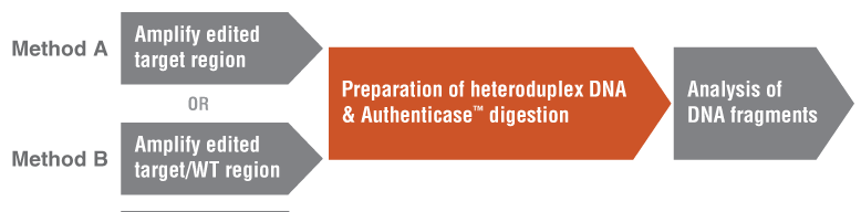
Note: Authenticase is ONLY USED in the second section of this workflow (Figure 2). NEB provides additional information and protocols to enable a more thorough workflow for mismatch detection during analysis of genome editing efficiency. These protocols were optimized and confirmed by NEB.
Two analytical methods are recommended to estimate the heterogeneity within a dsDNA pool generated by PCR amplification of an edited target region:
Method A is a conventional and popular setup (only requires amplifying DNA from edited cells) and provides a quick quantitative estimation (e.g., 53.1% mutation from a pool with 60% heterogeneous mutations (Case 1) and 27.9% for a pool with 60% homogeneous mutations (Case 2)).
Method B is a modified protocol (requires amplifying DNA from edited cells and wild type cells, respectively) that will provide a more accurate estimation of a pool with 60% heterogeneous mutation population (e.g., Case 1: 57.9% of Method B vs. 53.1% of Method A) including extreme cases where one type of specific mutation species comprises more than 50% of all the amplicons (as shown in Case 2: 47.7% of Method B vs. 27.9% of Method A).
Figure 1: Two methods for creating heteroduplex DNA for mismatch detection a assay
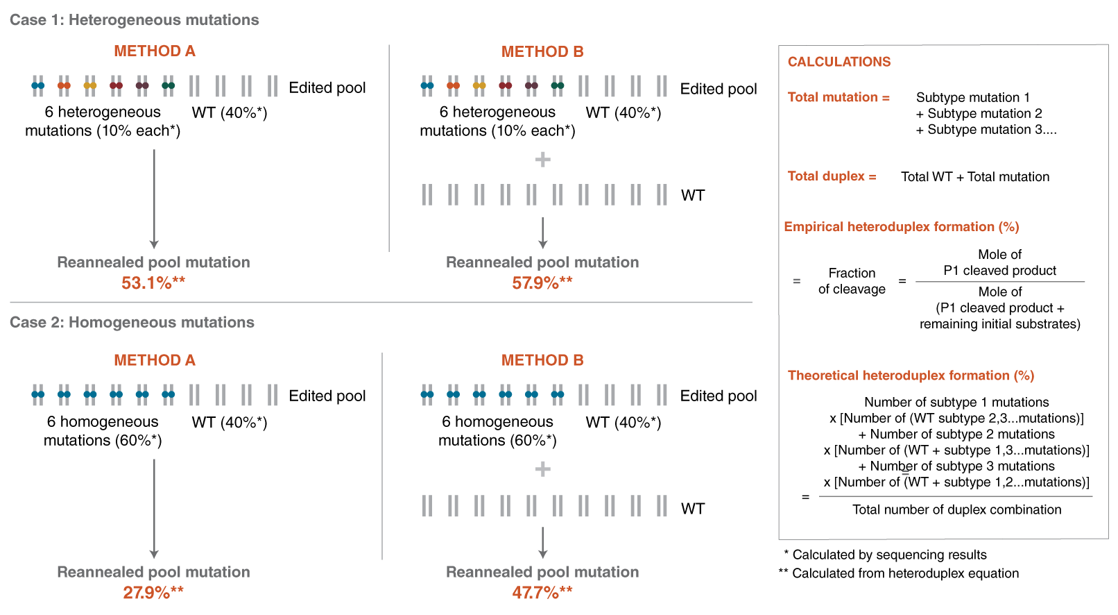
Figure 2: Mismatch Detection Assay Workflow
In practice, many users may choose to examine editing efficiency in target regions amplified from the edited cell population only (Method A). In cases where there is a dominant mutation (previously identified or suspected), having control gDNA from unedited cells can serve as a useful control and allow for an increased accurate calculation if Method B is followed. The following section provides guidance for amplification of both populations to enable this comparison.
1. Amplify target regions by PCR.
1.1. Thaw Q5 Hot Start High-Fidelity 2X Master Mix (purchased separately). Pulse-spin each component in microfuge prior to use.
1.2. Set up two 25 μl PCR reactions and use up to 500 ng of genomic DNA as templates. Reaction A is the experimental reaction with edited genomic DNA as template. Reaction B is the control reaction using gDNA from non-edited cells. Assemble the following reactions at room temperature:
|
REAGENT |
REACTION A |
REACTION B |
FINAL RXN. CONC. |
|---|---|---|---|
|
Q5 Hot Start High-Fidelity 2X Master Mix |
12.5 µl |
12.5 µl |
1X |
|
10 μM Forward Primer |
1.25 µl |
1.25 µl |
0.5 µM |
|
10 μM Reverse Primer |
1.25 µl |
1.25 µl |
0.5 µM |
|
Template DNA (edited genome) |
Variable |
|
0.5–500 ng genomic DNA** |
|
Template DNA (WT genome) |
|
Variable |
0.5–500 ng genomic DNA** |
|
Nuclease-free water |
to 25 µl |
to 25 µl |
|
* NEB recommends designing primers to produce amplicons around 700 bp with anticipated sizes of cleaved product around 450 and 250 bp, respectively, if a Bioanalyzer will be used at the end to analyze the genome editing efficiency.
** To use cell lysate directly in PCR, lyse cells in QuickExtract™ using 50 μl cells in each well of a 96-well plate (~40,000 cells) according to the manufacturers’ recommendation. Dilute the lysate 1:5 in TE and use 2.5 μl of the diluted lysate.
1.3. Gently mix the reaction. Collect all the liquid to the bottom of the tube with a brief spin. Transfer the tubes to a thermocycler and use the following conditions:
|
CYCLE STEP |
TEMP |
TIME |
CYCLES |
|---|---|---|---|
|
Initial Denaturation |
98°C |
2 minutes |
|
|
Denaturation |
98°C |
10 seconds |
35 |
|
Annealing |
50–72°C* |
5 seconds |
|
|
Extension (for 500–700 bp) |
72°C |
30 seconds |
|
|
Final Extension |
72°C |
2 minutes |
|
|
Hold |
4–10°C |
|
|
* Please visit tmcalculator.neb.com to determine correct annealing temperature.
2. Heteroduplex DNA formation and digestion with Authenticase.
The products of the PCR reaction must be denatured and re-annealed to allow formation of heteroduplexes between PCR products with and without mutations. A rapid qualitative analysis can be done by reannealing unpurified PCR amplicons followed by Authenticase digestion. An existing protocol can be slightly modified to include replacing T7 Endo I with Authenticase using a 42°C reaction temperature and incubation time of 15 minutes.
Optional: If the efficiency of genome editing will be calculated, NEB recommends purification of the reactions prior to fragment analysis. One can either use enzymatic treatment with Thermolabile Exonuclease I (NEB #M0568) and Quick CIP (NEB #M0525) to remove remaining primers and dNTPs or a spin column (e.g., Monarch PCR & DNA Cleanup Kit (5 μg) – NEB #T1030) to purify the annealed dsDNA.
Method A: PCR amplicons from genome of edited cells
This method only requires PCR amplicons from the genome of edited cells to proceed.
2A.1 DNA cleanup
It is critical to accurately quantitate DNA concentrations used as input for the mismatch detection assay (Step 2A.3.). To prepare DNA for accurate quantitation, PCR reactions can be cleaned up by an enzymatic method (Step 2A.1.1.) or a column method (Step 2A.1.2.) prior to preparation of heteroduplex DNA formation.
2A.1.1. Enzymatic cleanup method
2A.1.1.1. Measure the dsDNA concentration by Qubit® DNA quantification method.
2A.1.1.2. Prepare samples for enzymatic cleanup and heteroduplex DNA formation.
|
REAGENT |
REACTION |
|---|---|
|
PCR reaction (unpurified) |
18 µl |
|
Thermolabile Exonuclease I (NEB #M0568) |
1 µl |
|
Quick CIP (NEB #M0525) |
1 µl |
|
Total Volume |
20 µl |
2A.1.1.3. Briefly spin down all tubes and incubate reactions at 37°C for 4 minutes followed by 80°C for 1 minute. Proceed to Step 2A.2. directly to generate heteroduplex DNA.
2A.1.2. Column cleanup method
2A.1.2.1. Clean up PCR products using Monarch PCR & DNA Cleanup Kit (5 μg – NEB #T1030) with elution volume of 12 µl. Measure the dsDNA concentration.
2A.1.2.2. Prepare annealing reaction with cleanup DNA.
|
REAGENT |
REACTION |
|---|---|
|
Cleanup PCR amplicons (400 ng) |
1–16 μl |
|
5X Annealing Buffer |
4 μl |
|
Nuclease-free water |
to 20 µl |
2A.1.2.3 Go directly to Step 2A.2. to generate heteroduplex DNA.
2A.2. Heteroduplex DNA formation
2A.2.1. Generate heteroduplex DNA products in a thermocycler using the following program:
|
CYCLE STEP |
TEMP |
RAMP RATE |
TIME |
|---|---|---|---|
|
Initial Denaturation |
95°C |
|
3 minutes |
|
Annealing |
95–85°C |
2°C/second* |
|
|
85–25°C |
–0.1°C/second* |
|
|
|
Hold |
4°C |
|
|
* Alternatively, if a thermocycler is not available with these ramp speeds, the sample can be heated to 95°C for 10 minutes and then allowed to cool slowly to room temperature.
2A.3. Digestion of heteroduplex DNA with Authenticase
2A.3.1. Digest heteroduplex DNA with Authenticase as follows:
|
REAGENT |
ENZYMATIC-CLEANUP AMPLICONS |
COLUMN-PURIFIED AMPLICONS |
||
|---|---|---|---|---|
|
Reaction |
Negative Control |
Reaction |
Negative Control |
|
|
Annealed PCR amplicons (~200 ng) |
1–12 μl* |
1–12 µl* |
10 μl |
10 µl |
|
10X Reaction Buffer |
2 µl |
2 µl |
2 µl |
2 µl |
|
Authenticase |
1 μl |
0 µl |
1 μl |
0 µl |
|
Nuclease-free water |
16–5 µl |
17–6 µl |
7 µl |
8 µl |
|
Total Volume |
20 µl |
20 µl |
20 µl |
20 µl |
* The digestion reaction conditions have been optimized for up to 6 μl of the unpurified enzyme-treated Q5 Master Mix PCR reaction or 12 µl of unpurified OneTaqPCR reaction containing up to 200 ng of amplified DNA. Increased amounts of PCR reaction and/or DNA may lead to inaccurate estimates of editing efficiencies.
2A.3.2. Mix well and briefly spin. Incubate each reaction at 42°C for 15 minutes. Stop the reaction with 1.7 µl of 150 mM EDTA. Proceed with fragment analysis or store at –20°C until ready.
Method B: PCR amplicons from genome of edited cells and WT cells
This method requires both PCR amplicons from the genome of edited cells and WT cells to proceed.
2B.1. DNA cleanup
Amount of DNA concentration is essential in the mismatch detection assay. PCR reactions can be cleaned up by an enzymatic method (Step 2B.1.1.) or a column method (Step 2B.1.2.) prior to preparation of heteroduplex DNA formation.
2B.1.1. Enzymatic cleanup method
2B.1.1.1. Measure the PCR amplicons concentration by Qubit® DNA quantification method.
2B.1.1.2. Prepare samples for enzymatic cleanup and heteroduplex DNA formation.
|
REAGENT |
REACTION A |
REACTION B |
|---|---|---|
|
PCR amplicons from edited genome |
18 µl |
|
|
PCR amplicons from WT genome |
|
18 µl |
|
Thermolabile Exonuclease I (NEB #M0568) |
1 µl |
1 μl |
|
Quick CIP (NEB #M0525) |
1 µl |
1 µl |
|
Nuclease-free water |
to 20 µl |
to 20 µl |
2B.1.1.3. Briefly spin down all tubes and incubate reactions at 37°C for 4 minutes followed by 80°C for 1 minute.
2B.1.1.4. Prepare samples to make heteroduplex DNA by mixing 200 ng of reaction A (from edited gDNA template) and 200 ng of reaction B (from WT gDNA template). Assemble the reaction as follows and go to step 2B.2.:
|
REAGENT |
REACTION |
|---|---|
|
Enzymatic cleanup PCR samples from edited cells (200 ng) |
X μl |
|
Enzymatic cleanup PCR samples from WT cells (200 ng) |
Y μl |
|
Total Volume |
X + Y µl |
2B.1.1.5 Go to step 2B.2. to generate heteroduplex DNA.
2B.1.2. Column cleanup method
2B.1.2.1. Clean up PCR products using Monarch PCR & DNA Cleanup Kit (5 μg – NEB #T1030) with elution volume of 12 µl. Measure the dsDNA concentration.
2B.1.2.2. Prepare annealing reaction with cleaned up DNA.
|
REAGENT |
REACTION |
|---|---|
|
Column cleanup PCR samples from edited cells (200 ng) |
X µl |
|
Column cleanup PCR samples from edited cells (200 ng) |
Y µl |
|
5X Annealing Buffer |
4 µl |
|
Nuclease-free water |
to 20 µl |
2B.1.2.3. Go to step 2B.2. to generate heteroduplex DNA.
2B.2. Heteroduplex DNA formation
2B.2.1. Generate heteroduplex DNA products in a thermocycler using the following program:
|
CYCLE STEP |
TEMP |
RAMP RATE |
TIME |
|---|---|---|---|
|
Initial Denaturation |
95°C |
|
2 minutes |
|
Annealing |
95–85°C |
2°C/second* |
|
|
85–25°C |
–0.1°C/second* |
|
|
|
Hold |
4°C |
|
|
* Alternatively, if a thermocycler is not available with these ramp speeds, the sample can be heated to 95°C for 10 minutes and then allowed to cool slowly to room temperature.
2B.3. Digestion of heteroduplex DNA with Authenticase
2B.3.1. Digest heteroduplex DNA with Authenticase as follows:
|
REAGENT |
ENZYMATIC-CLEANUP AMPLICONS |
COLUMN-PURIFIED AMPLICONS |
||
|---|---|---|---|---|
|
Reaction |
Negative Control |
Reaction |
Negative Control |
|
|
Annealed PCR amplicons (~200 ng) |
(X + Y)/2 μl* |
|
10 μl |
|
|
Annealed PCR product (~200 ng) |
|
(X + Y)/2 μl* |
|
10 µl |
|
10X Reaction Buffer |
2 µl |
2 µl |
2 µl |
2 µl |
|
Authenticase |
1 μl |
0 µl |
1 μl |
0 µl |
|
Nuclease-free water |
17 – (X + Y)/2 µl |
18 – (X + Y)/2 µl |
7 µl |
8 µl |
|
Total Volume |
20 µl |
20 µl |
20 µl |
20 µl |
* The digestion reaction conditions have been optimized for up to 6 μl of the unpurified enzyme-treated Q5 Master Mix PCR reaction or 12 µl of unpurified OneTaq PCR reaction containing up to 200 ng of amplified DNA. Increased amounts of PCR reaction and/or DNA may lead to inaccurate estimates of editing efficiencies.
2B.3.2. Mix well and briefly spin. Incubate each reaction at 42°C for 15 minutes. Stop the reaction with 1.7 µl of 150 mM EDTA. Proceed with fragment analysis or store at – 20°C until ready.
3. Analyze DNA fragments.
3.1 Gel or Fragment Analysis
3.1.1. Add 4 μl of Gel Loading Dye, Purple (6X, NEB #B7024) to the reaction and run on a 2% agarose gel stained with ethidium bromide.
3.1.2. Run the included DNA ladder or an appropriate DNA size marker alongside the sample for reference. Alternatively, samples can be analyzed using a fragment analyzer (e.g., Agilent Bioanalyzer or Advanced Analytical Technologies, Inc. (AATI) Fragment Analyzer). For the Agilent Bioanalyzer, remove 2 µl of enzyme-treated sample and mix 8 µl of water.
3.1.3. Analyze 1 μl of the 5X diluted sample on a high sensitivity Agilent DNA chip. This allows for detection of mutation populations down to 1 out of 80 copies based on 700 bp PCR amplicon design with cleaved product sizes of 450 bp and 250 bp. For the AATI Fragment Analyzer, 2 μl of the reaction can be used with the Standard Sensitivity NGS Fragment Analysis Kit (AATI Cat# DNF-473).
3.2 Efficiency Calculation
3.2.1. Calculate the estimated % modification using the following formula:
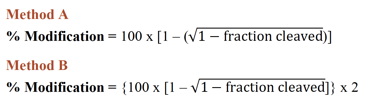
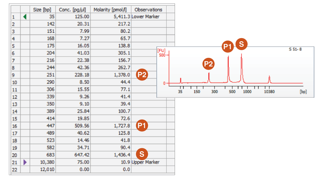
When calculating % modification for reactions with the control template where the starting material (S) is known, the equation (100 x fraction cleaved) can be used, where fraction cleaved (also known as % heteroduplex in Figure 4) = molarity of digested products (P1 and P2*)/(molarity of digested products + molarity of undigested band).**
* Note: P2 does not factor into calculation.
** Reference: Sentmanat, M.F., et al. (2018) Sci Rep. 8(1), 888.
Example of Calculation of Data from Bioanalyzer Results
An example of % efficiency sample data:
![]()